


A retinal detachment is a serious medical emergency. While painless, it can quickly lead to blindness if not treated.
Luckily, there are treatments available to save your retina and your vision. However, you need to know how to recognize if you could have a retinal tear or detachment to know when to get treatment. It’s also important to be aware of your risk for retinal detachment.
Keep reading to learn more about what a retinal detachment is, the symptoms, who is at risk for it, and how to treat them if you have one.
What is a Retinal Detachment?
Your retina is the thin membrane on the back of your eye that contains photoreceptor cells. These cells translate the light that passes through your eye into impulses sent to your brain.
When your brain receives these impulses, it processes them into images you can see. Without your retina, you wouldn’t be able to see at all.
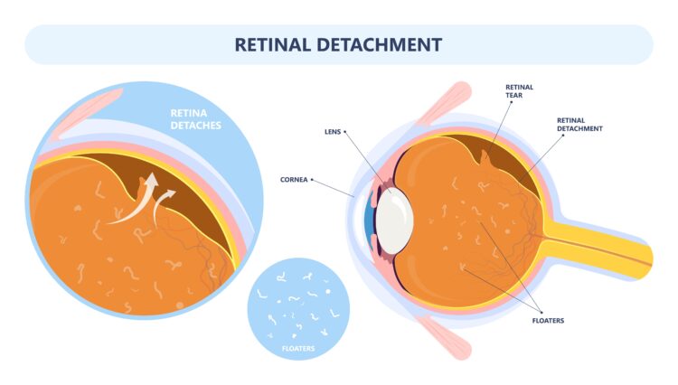
A retinal detachment occurs when the retina detaches from the eye. If the retina pulls away from its position in the eye, it cuts the retina off from blood vessels, depriving it of oxygen.
If left unattached for too long, the photoreceptor cells on the retina will die, leaving you unable to see even if you can have the retina reattached.
Thinning of the membrane causes retinal detachments. This makes the membrane more susceptible to tears. A retinal tear often leads to a retinal detachment, making it easier for the retina to pull away from the back of your eye.
You can also experience a retinal detachment when fluid builds up in the eye or scar tissue in the eye. The scar tissue may come from an eye injury or a previous eye surgery. No matter what causes a retinal detachment, it is a medical emergency requiring immediate attention that permanently damages your vision if left untreated.
Symptoms of a Retinal Detachment
Retinal tears and detachments are painless, but they do cause visual symptoms. These symptoms include:

- Seeing flashes of light
- Noticing floaters
- Darkening of your peripheral vision
- Seeing a shadow that covers part of your vision
If you experience these symptoms, seek immediate medical attention. If you can’t get an appointment immediately with your eye doctor at St. Luke’s at The Villages, go to the emergency room.
Remember to keep in mind that retinal detachment is much more common in older patients. If you’re at an increased risk of developing a retinal detachment, you may already have some floaters in your vision.
But if you suddenly notice many more floaters, you could have a retinal tear or detachment and should seek medical attention.
Risk Factors of a Retinal Detachment
Retinal detachment is rare, but it’s more common in people who include the following:

- Are very nearsighted
- Are over 50
- Have lattice degeneration, which is the thinning of the retina
- Have a history of retinal tears
- Have had an eye injury
- Have a family history of retinal detachment
- Have had a previous eye surgery
If any of these factors describe you, you should be vigilant about the possibility of retinal tears and detachments. You should also see your eye doctor regularly. Regular eye exams with your eye doctor ensure they can spot the early signs of lattice degeneration and other age-related eye conditions that may affect your vision.
Treating a Retinal Detachment
You need immediate treatment after a diagnosis of having a retinal detachment. There are several options for treatment.
Your eye doctor will decide on a treatment based on the severity of the retinal detachment. Sometimes, if you only have a tear and the retina hasn’t yet detached, treatment can include one of two simple procedures:
Photocoagulation
During photocoagulation, a laser burns a tear or hole in the retina, creating scar tissue that holds the retina together. By creating scar tissue, a surgeon can reattach the retina to the back of the eye.
Cryopexy
Cryopexy is a procedure similar to photocoagulation. It uses a freezing probe on the outside of your eye over a hole or a tear to create scar tissue to repair the tear. The scarring created helps keep the retina in place.
Retinal detachments require somewhat more extensive procedures, although which one depends on how severe the detachment is. These procedures include:
Pneumatic Retinopexy
Pneumatic retinopexy is a procedure that repairs minor detachments. Your ophthalmologist can perform it in-office. They will place a gas bubble in the eye to push the retina back into place.
The gas bubble works as a splint in the eye. After pneumatic retinopexy, patients must keep their heads positioned in a certain way to keep the gas bubble intact and in the correct spot. The gas bubble goes away on its own after a few weeks while the eye heals.
Scleral Buckling
Scleral buckling is a procedure that treats larger retinal detachments. The technique involves putting a band on the outside of the eye (the sclera) to push it back into the retina in the back of your eye.
You may have a retinopexy or cryopexy paired with scleral buckling to simultaneously fix a tear and detachment. You may also have it performed in combination with a vitrectomy.
Vitrectomy

Vitrectomy treats severe retinal detachments. Patients require surgery to access the inside of the eye and remove part of the vitreous, the gel that fills your eye.
By removing part of it, there’s less pressure on the retina. Having less pressure on the retina allows it to return to its correct position, usually with a gas bubble. Your surgeon may also choose to perform a cryopexy or retinopexy with a vitrectomy to fix any tears.
These treatments can save your vision. If you experience any retinal detachment symptoms, seek attention right away. Without prompt diagnosis and treatment, you can go blind.
Take charge of your vision by scheduling an appointment at St. Luke’s at The Villages in Lake Sumter Landing, FL, today!


