Our retina team is highly experienced in treating vitreoretinal diseases including diabetic retinopathy, macular degeneration, retinal detachment, macular hole, and other vitreoretinal disorders. As part of our mission to provide patients with life-changing vision, St. Luke’s at The Villages is proud to offer retina eye care.
Our retina specialist, Dr. Remington Horesh, is a board-certified, fellowship-trained vitreoretinal surgeon who specializes in the medical and surgical treatment of retinal disorders. Dr. Horesh, along with the rest of our retina team, is here to take care of your retina-related concerns and conditions and to get you one step closer to preserving your vision.
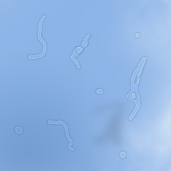
Floaters in your vision
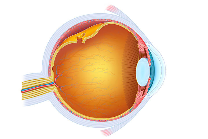
The space between the intraocular lens and the retina is filled with a clear, gel-like substance called vitreous. In a newborn, the vitreous has a solid but clear consistency and is firmly attached to the retina. With natural progression over time, the vitreous degenerates, liquefies and may separate from the retina, termed, a posterior vitreous detachment (PVD). This is a very common process and typically harmless. During this process, the vitreous loses its optical clarity and may result in hazy, cloudy vision, with or without identifiable floaters. Floaters are caused by tiny clumps of denatured gel and cells that cast bothersome shadows onto the retina.
Occasionally, floaters can be accompanied by flashes, which represent traction by the vitreous on the retinal tissue. In most cases this traction resolves in an uncomplicated fashion but can represent varying sight-threatening retinal conditions and therefore a dilated exam by an eye care professional is warranted if these symptoms are present.
Common Floater Symptoms
– Black spots or “spider webs” that appear to float in the vision.
– Dark spots that move around or remain suspended in one place.
– “Cloud-like” haze that can float in the vision.
Treatment for Floaters
While floaters never truly disappear from the eye on their own, many people will become less symptomatic over time as they “settle” and as the brain learns to ignore them. Some individuals will tolerate them, and some choose to live with them, but you don’t have to. If you are symptomatic of floaters interfering with your clarity of vision and affecting your daily life activities, there is an effective solution. With advancing technology, a small-gauge vitrectomy procedure, performed by a vitreoretinal surgeon in the outpatient setting, can remove all the floaters within your eye, restoring clarity and relieving you from those visually disabling vitreous floaters. With a high success rate and great safety profile similar to modern-day cataract surgery, floaters can be effectively treated and your quality of life improved.
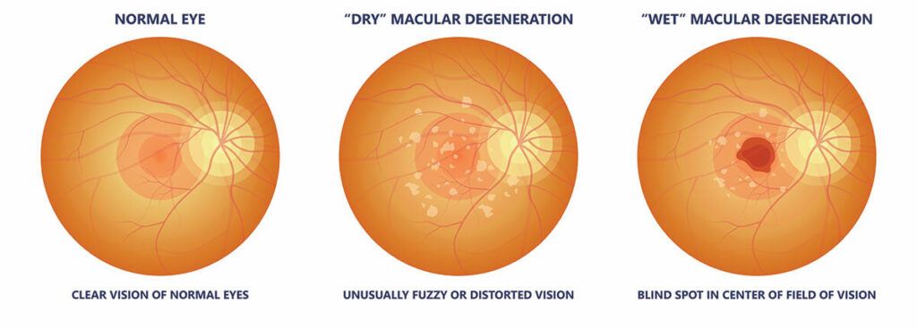
Age-Related Macular Degeneration (AMD) is an age-related, degenerative issue with the retina. It occurs when the center of the retina is damaged, resulting in central vision problems. It happens when the macula— the part of the eye located near the center of the retina that controls sharp, straight-ahead vision—is damaged due to aging. The damage to the central vision leads to difficulty reading, seeing fine details, and recognizing faces. AMD does not impact one’s peripheral (side) vision, but rather is a disease that affects the center of one’s vision.
AMD is a very common condition in people over the age of 60 and is a leading cause of vision loss for older adults. It often affects both eyes, however, one eye may be more affected than the other. The severity of the disease varies greatly, as some people who have the disease may not notice any changes at all. It is a condition that develops very slowly in some people, and much faster in others.
There are two types of AMD: Dry and Wet.
Dry AMD
The majority of people with AMD have dry AMD, also known as atrophic AMD. Dry AMD makes up about 90% of the cases of AMD. With this type of AMD, the macula thins as one ages. Those with this type of AMD may have yellow deposits, known as drusen, in the macula.
A few small drusen may not have an impact on your vision, but as they get bigger and become more numerous, they can distort or dim your vision and can cause central vision loss.
Dry AMD occurs in 3 stages: early, intermediate, and late. It usually slowly progresses over several years.
Wet AMD
Wet AMD, also known as neovascular AMD, is a less common and more serious type of late AMD. Only about 10% of people with macular degeneration have this form. In this type of AMD, abnormal blood vessels grow under the macula, bleed, and leak fluid into the retina. This can cause vision to become distorted, can create blind spots, and lead to a loss of central vision. These blood vessels are like weeds growing through cracks and eventually, they form a scar which can lead to permanent loss of central vision. Any stage of dry AMD can turn into wet AMD, but wet AMD is always the late stage of this condition.
Compared to dry AMD, wet AMD progresses much quicker and can cause patients to experience a more significant loss of vision. However, if caught early enough, there are treatment options that can stabilize a patient’s vision and lower the risk of experiencing further vision loss.
Treatment Options For AMD
Dry AMD
There are currently no treatments for dry AMD, but there are steps you can take, such as consuming specially formulated vitamins, to decrease the risk of the condition progressing into its most advanced form.
Wet AMD
To treat wet AMD, anti-VEGF medications are commonly used. This form of treatment is injected directly into the eye and works to reduce the number of abnormal blood vessels in the retina.
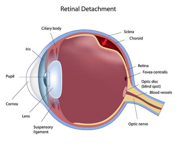
Retinal Detachment
The retina is a light-sensitive membrane that is located at the back of the eye. When light passes through the eye, the lens focuses an image on the retina. The retina then converts that image into signals that are sent to your brain through the optic nerve. The retina works with the lens, cornea, and other parts of the eye and brain in order to produce normal vision.
What causes retinal detachment?
When the retina separates from the back of the eye, retinal detachment occurs. This detachment can cause either a partial or a total loss of vision, all depending on how much of the retina is detached. When the retina becomes detached, it is pulled from the tiny blood vessels (known as choroids) that supply it with both oxygen and nutrients. The retinal cells may become severely deprived of oxygen, which increases the risk of suffering from permanent loss of vision. The longer these cells are separated from the retina, the more likely a person is to experience a permanent loss of vision. Retinal detachment is a medical emergency—contact St. Luke’s at The Villages immediately if you begin to experience any sudden vision changes.
Potential risk factors for retinal detachment include:
Posterior vitreous detachment, which is common in older adults
Extreme nearsightedness, which is known to cause more strain on the eye
A family history of retinal detachment
Prior history of retinal detachment or tear
A history of glaucoma (an eye disease related to eye pressure)
A history of weak area in your retina (known as lattice degeneration and has been seen by your eye doctor in previous exams)
Nearsightedness (requiring glasses to see far away)
Notify Dr. Remington Horesh immediately if you notice any of the above.
There is no pain related to retinal detachment, but there is usually some discomfort and symptoms prior to the retina becoming detached.
Primary symptoms of the condition include:
Blurred vision
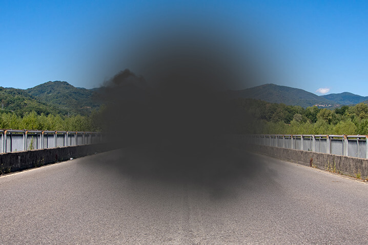
Partial vision loss, which makes it seem as though a curtain has been pulled across your field of vision, giving off a dark, shadowing effect
Sudden flashes of light that appear in your peripheral vision (when looking to the side)
Suddenly seeing floaters, which are tiny pieces of debris that look like black flecks or strings floating before your eye
An increase in flashes or floaters
There Are Three Types of Retinal Detachment
Rhegmatogenous Retinal Detachment
Rhegmatogenous retinal detachment means that one has a tear or hole in their retina. The tear or hole allows for fluid from inside the eye to slip through the opening and get behind the retina. The fluid separates the retina from the retinal pigment epithelium, the membrane that provides the retina with nourishment and oxygen, which causes the retina to detach. Rhegmatogenous retinal detachment is the most common type of retinal detachment.
Tractional Retinal Detachment
When scar tissue on the retina’s surface tightens, it causes the retina to pull away from the back of the eyes, leading to tractional retinal detachment. This type of detachment is less common than other types, and typically affects people with diabetes mellitus. When diabetes mellitus is inadequately controlled or improperly taken care of, it can lead to issues with the retinal vascular system. This vascular damage can eventually lead to the accumulation of scar tissue in the eye that could cause retinal detachment.
Exudative Detachment
In cases of exudative detachment, there are no tears or breaks in the retina. Rather, retinal diseases such as the following cause this type of detachment:
An inflammatory disorder that causes fluid to build up behind your retina
Cancer behind your retina
Coats’ disease, which causes abnormal development in the blood vessels leading to leaking proteins building up behind the retina
Treatment for Retinal Detachment
In most cases, surgery is the necessary form of treatment to repair a detached retina. However, for minor retinal detachments or tears, a simple procedure may be done in our surgical center at St Luke’s at The Villages Sumter Landing. Having said that, surgery is almost always used to repair a retinal tear, hole, or detachment, and there are several techniques available:
Photocoagulation-If there is a hole or tear in the retina but the retina is still attached, your doctor may use a laser to perform a procedure called photocoagulation. The laser burns around the tear site, and the resulting scarring attaches your retina to the back of your eye.
Cryopexy- Cryopexy, which uses intense cold to freeze an area of your eye, is another option. In this treatment, your doctor applies a freezing probe to the outside of your eye in the area over the retinal tear site, and the resulting scarring helps keep your retina in place.
Pneumatic Retinopexy– Pneumatic retinopexy is an option to repair minor detachments that can be done in the office. In this procedure, your doctor places a gas bubble inside your eye to help your retina move back into place up against the eye’s wall. The gas bubble acts as a splint. After this procedure, it is imperative to keep your head positioned in a specific way for a few days to allow the bubble to remain in the right spot. The gas bubble goes away on its own over the course of a few weeks as your eye heals.
Scleral buckling– Your doctor may recommend scleral buckling, which involves placing a band around the outside of the eye to push the wall of the eye into the retina, getting it back into place for proper healing. Scleral buckling may be done in combination with a vitrectomy, and a cryopexy or retinopexy is performed during the scleral buckle procedure.
Vitrectomy Surgery- Another treatment option is a vitrectomy, which is used for larger tears. This procedure involves anesthesia and is often performed in an operating room as an outpatient procedure. Here, your doctor will use small tools to remove vitreous, the gel-like fluid that fills your eyes, and abnormal vascular or scar tissue from your retina. The doctor will then put your retina back into its proper place, which is usually done with a gas bubble. A Cryopexy or retinopexy is performed during the vitrectomy procedure.
Retina Eye Care Doctor
Remington Horesh, DO
“The field of Vitreoretinal Surgery is complexly detailed, innovative, and vital to preserving and restoring your vision. I am enthusiastic to be a part of the expert team at St. Luke’s as your vitreoretinal surgeon. I look forward to providing my patients in The Villages with the most personalized and advanced retinal care in the world.”
Remington Horesh, DO
In addition to his formal training, Dr. Horesh’s past and present experiences at the forefront of therapeutic advancements in retinal disease have been remarkably advantageous to his knowledge and expertise in the field. He has served as a treating physician in large-scale clinical trials aimed at discovering new and effective treatments for age-related macular degeneration.
If you or someone you know is struggling with AMD or any retinal-related eye diseases and needs an expert opinion, call 352-775-0080 to schedule a consultation.
We proudly serve patients at our eye clinics in Tarpon Springs, Tampa, St. Petersburg, Clearwater, Spring Hill, Wesley Chapel and in The Villages at Lake Sumter Landing and Brownwood. Our philosophy is to treat those we serve as though they are a member of our own families. From the first phone call or email to the follow-up visit and beyond, we’re here to serve you. Our goal is to help preserve and optimize your vision.